SonoSK
An open-source Python library for medical ultrasound. Design a probe, place it in front of a tissue phantom, and get the B-mode image, or heat a target and see the temperature, the thermal dose and the lesion left behind.
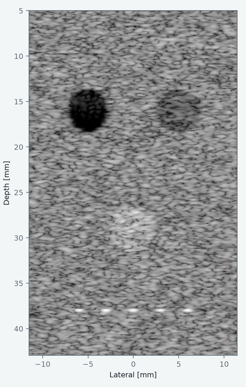
What it simulates
- Transducers
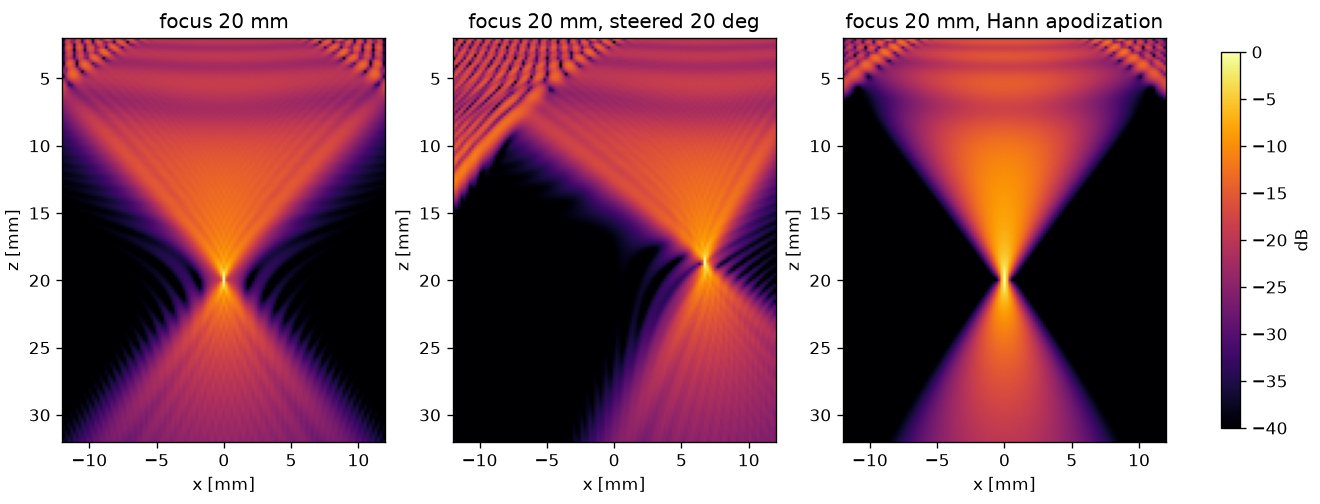
- Flat and focused elements, annular arrays, linear and phased arrays with an elevation lens, convex and endocavity arrays, matrix arrays and line-focus sources, with focusing, steering and apodization.
- Pressure fields
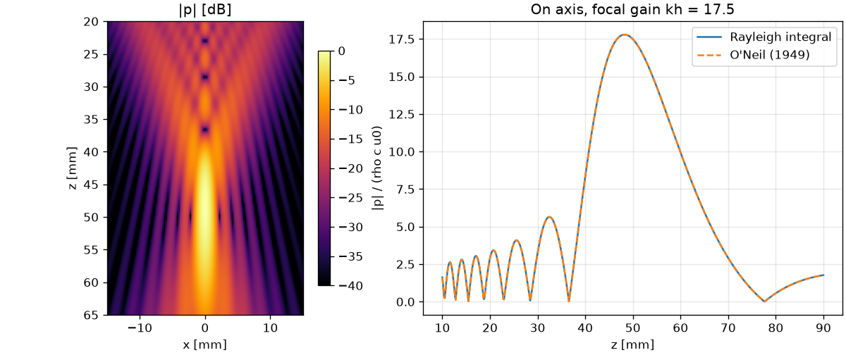
- Rayleigh integral in an attenuating medium and the angular spectrum method, on lines, planes and volumes.
- Imaging
- Pulse-echo signals from scatterer phantoms, plane, diverging and focused transmits, delay-and-sum beamforming and compounding.
- Therapy
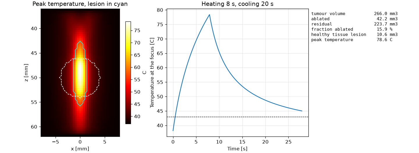
- Heat from the field, the Pennes bioheat equation in 1, 2 and 3 dimensions, CEM43 dose, lesion maps, and the ablated and residual target.
- Probe design
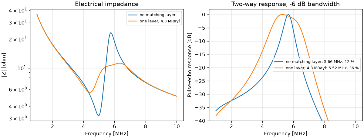
- Mason model of a piezoelectric element with backing and matching layers: impedance, transmit and pulse-echo response.
- Safety and quality
- Mechanical index, derated intensities and FDA limits; resolution, contrast, CNR, gCNR and speckle statistics.
Figures made with SonoSK
A first simulation
import sonosk as sk
from sonosk import field, transducer

probe = transducer.linear_array(128, pitch=0.3e-3, height=5e-3, elev_focus=20e-3)
probe.focus((0, 0, 20e-3), c=1540.0)

grid = sk.Grid.centered((121, 151), 0.2e-3, depth_start=2e-3)
p = field.rayleigh_grid(probe, grid, f=5e6, c=1540.0, alpha0=0.5)
The getting started page continues from here to a phantom, an image, its quality measures and a heating exposure. Each solver is checked against a closed-form result; the comparisons are on the validation page, and the plan for tissue data and organ phantoms is on the roadmap.
Citing SonoSK
Kooklan S. SonoSK: medical ultrasound simulation in Python. Zenodo, 2026. doi:10.5281/zenodo.23517377
This DOI always resolves to the latest release, and every release has its own DOI as well. BibTeX and the DOI of each version are on the citing page.