Anatomical phantoms
sonosk.organs builds phantoms of the breast, prostate, uterus and facial
skin on 1-, 2- or 3-D grids. Every region takes its properties from
sonosk.tissues, layer thicknesses default to published
means, and the interfaces undulate with smooth random profiles, so every
seed gives a different but plausible anatomy.
import sonosk as sk
from sonosk import organs

g = sk.Grid.centered((401, 401), 0.1e-3, depth_start=0.0)     # x, z in metres
ph = organs.breast(g, lesions=[organs.Cyst((-11e-3, 0, 13e-3), a=3e-3, b=2.4e-3),
                               organs.Fibroadenoma((0, 0, 14.5e-3)),
                               organs.Carcinoma((11e-3, 0, 16e-3), radius=3.2e-3)],
                   rng=4)
ph.medium          # Medium with labels and materials
ph.targets         # names of the lesions
ph.description     # sizes used
ph.sources()       # citations of all tissue values
The depth axis points away from the probe, which sits at z = 0. A 3-D grid (x, y, z) gives a 3-D phantom with the same call.
Lesions
Class |
Shape |
|---|---|
|
ellipsoid, anechoic |
|
oval, wider than tall, smooth lobulated margin |
|
irregular, spiculated, taller than wide |
|
generic irregular mass of any tissue, for example a fibroid or a prostate tumour |
|
small bright inclusion with high attenuation |
The shapes follow the BI-RADS descriptors of benign and suspicious masses.
Each lesion can be given a label, its name in the medium.
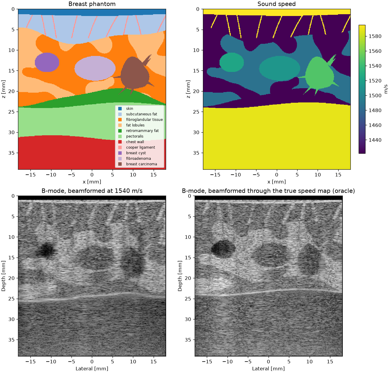
Breast
organs.breast(grid, lesions, skin=1.45e-3, fat=5e-3, gland=15e-3, retro_fat=2e-3, muscle=8e-3, density=0.6, ligaments=True, ribs=False)
Skin, subcutaneous fat with Cooper’s ligaments, fibroglandular tissue
mixed with fat lobules (density is the glandular fraction), retromammary
fat, pectoralis and chest wall. The default skin thickness is the mean of
1.44-1.45 mm measured by dedicated breast CT (Shi et al. 2013; Huang et al.
2008).
Prostate
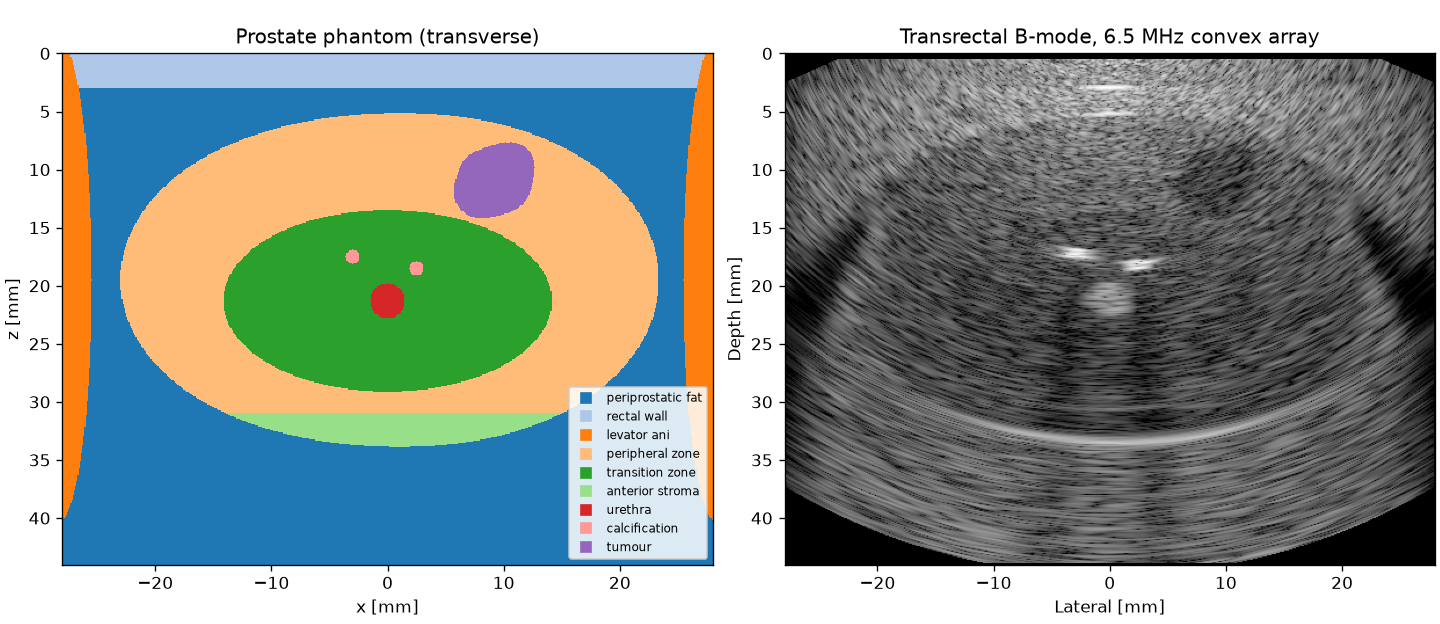
organs.prostate(grid, tumors, calcifications, rectal_wall=3e-3, fat=1.5e-3, size=(45e-3, 35e-3, 30e-3), tz_fraction=0.45)
A transverse section seen from the rectum: rectal wall, periprostatic fat, peripheral zone facing the probe, transition zone around the urethra, anterior fibromuscular stroma and levator ani. The default gland volume is about 25 mL, near the mean of 26.9 mL of patients treated by transrectal HIFU (Pichardo et al. 2008); the peripheral zone is about 9 mm thick behind the transition zone, inside the 1.6-15 mm range measured by MRI (Sellers et al. 2022).
Uterus
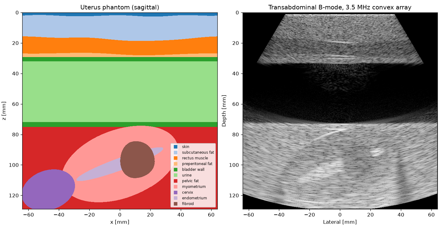
organs.uterus(grid, fibroids, approach="transabdominal", skin=2e-3, fat=15e-3, muscle=10e-3, bladder=40e-3, size=(80e-3, 50e-3, 45e-3), tilt=20 deg, endometrium=8e-3)
A sagittal section with skin, subcutaneous fat, rectus muscle, a full
bladder (wall 3 mm, Hakenberg et al. 2000) and an anteverted uterus with
myometrium, endometrium and cervix. The default uterus lies within the
mean sizes measured by ultrasound, 72.8 x 42.8 x 32.4 mm in nulliparous to
90.8 x 51.7 x 43.0 mm in multiparous women (Esmaelzadeh et al. 2004).
approach="transvaginal" removes the abdominal wall and the bladder.
Face
organs.face(grid, epidermis=0.1e-3, dermis=1.3e-3, fat=2.6e-3, smas=1.0e-3, muscle=8e-3, bone=False)
Epidermis, dermis, superficial fat, SMAS and facial muscle for aesthetic focused ultrasound. The full skin thickness of 1.4 mm lies in the 1.31-1.64 mm range measured over eight facial sites (Jeong et al. 2023), and the SMAS starts at 4.0 mm, the depth reached by the 4.5 mm micro-focused transducer (Chaves Bellote and Miot 2021).
Random structure
phantom.random_field, phantom.wavy_below, phantom.irregular and
phantom.curve are the building blocks behind the organs and can be used
for new ones. phantom.fluctuations(medium, level) adds sub-resolution
variations of density and speed whose strength follows the echo level of
each tissue; they make the speckle of full-wave images.
References
Chaves Bellote T.P., Miot H.A. Microfocused ultrasound with visualization for face slimming: preliminary results in four women. Clin. Cosmet. Investig. Dermatol. 14:1613-1619, 2021.
Esmaelzadeh S., Rezaei N., HajiAhmadi M. Normal uterine size in women of reproductive age in northern Islamic Republic of Iran. East. Mediterr. Health J. 10(3):437-441, 2004.
Hakenberg O.W., Linne C., Manseck A., Wirth M.P. Bladder wall thickness in normal adults and men with mild lower urinary tract symptoms and benign prostatic enlargement. Neurourol. Urodyn. 19(5):585-593, 2000.
Huang S.Y., Boone J.M., Yang K., Kwan A.L., Packard N.J. The effect of skin thickness determined using breast CT on mammographic dosimetry. Med. Phys. 35(4):1199-1206, 2008.
Jeong K.M., Seo J.Y., Kim A., et al. Ultrasonographic analysis of facial skin thickness in relation to age, site, sex, and body mass index. Skin Res. Technol. 29(8):e13426, 2023.
Pichardo S., Gelet A., Curiel L., Chesnais S., Chapelon J.Y. New integrated imaging high intensity focused ultrasound probe for transrectal prostate cancer treatment. Ultrasound Med. Biol. 34(7):1105-1116, 2008.
Sellers J., Wagstaff R., Helo N., de Riese W.T.W. Association between prostate size and MRI determined quantitative prostate zonal measurements. Res. Rep. Urol. 14:265-274, 2022.
Shi L., Vedantham S., Karellas A., O’Connell A.M. Skin thickness measurements using high-resolution flat-panel cone-beam dedicated breast CT. Med. Phys. 40(3):031913, 2013.